|
|
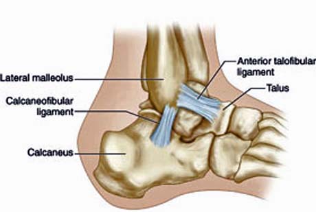
Calcaneofibular ligamentCalcaneofibular ligament DefinitionThe Calcaneofibular ligament is a rounded and narrow cord that runs to the lateral calcaneus surface from the apex of the fibula’s malleolus. It is 3 mm thick, 5 mm wide and 2 cm long.
This is a section of the lateral collateral ligament that contradicts the subtalar joint hyperinversion, as can be seen in a common form of ankle sprain. The brevis muscles and the tendons of the fibularis longus cover it. It is known by other terms like Ligamentum calcaneofibulare (Latin) and Ligament calcanéofibulaire (French). Calcaneofibular ligament Function
The ligament connects the calcaneus (heel) and talus bones of the foot. It is primarily responsible for controlling inversion. Inversion means turning of the foot sideways, so that the bottom of foot turns towards the contrary foot. The Calcaneofibular ligament keeps both the subtalar joint and the ankle stable.
Calcaneofibular ligament Injury
Damage to the ligament results once the foot is twisted too much when the toes point to the shin in an upwards fashion. The damage can be diagnosed by doctors with a talar tilt test. During this test, the patient has to sit on a bench and keep the foot slightly angled or flat. The leg is held over the ankle and the foot is manipulated to ensure inversion. In case of pain, the Calcaneofibular ligament is regarded by doctors as the underlying cause.
Calcaneofibular ligament Injury SymptomsThe signs and symptoms associated with Calcaneofibular ligament Injury include the following:
Calcaneofibular Ligament Tear
When the calcaneofibular ligament is torn, it is generally injured along with tears in the anterior talofibular ligament. Injuries in the ligaments at the ankle are seen reliably with MR (Magnetic Resonance), which manifests in the form of discontinuity or abnormal laxity in the impacted ligament. It can also manifest as edema and thickening of soft tissue around the ligament, during partial tearing. Chronic spraining of ligaments can be seen as structures that have been thickened abnormally, without the related edema (6a).
Impingement of soft tissue can result from injury to the lateral ligament in the ankle. It can lead to chronic Calcaneofibular Ligament pain with mechanical signs and symptoms. The soft tissue impingement sites typically include syndesmotic, anterolateral and posterior, of which anterolateral impingement is the commonest. Calcaneofibular Ligament MRI helps in the reliable visualization of the pathology at the ankle and the anatomy of the ligament. It is being increasingly used in patients after sprains in the lateral ankle. With the ongoing evolution of surgical techniques and indications for the reconstruction of lateral ligaments, the use and significance of MRI in preoperative assessment is only likely to increase. Calcaneofibular ligament Pictures
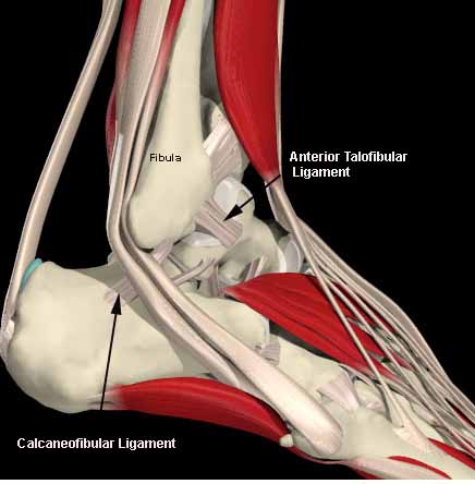
Check out the following Calcaneofibular ligament images and diagrams to know more about the structure.
|

|
|