|
|
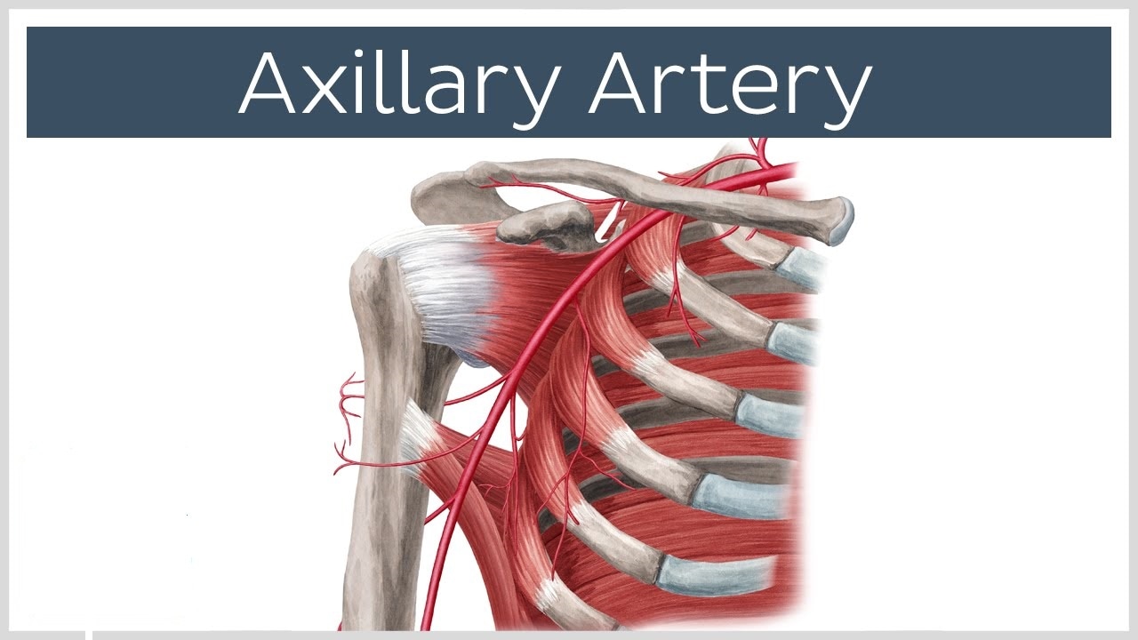
Axillary ArteryAxillary Artery Definition
The axillary artery is a prolongation or extension of the subclavian. It is known as Arteria axillaris in Latin and as Artère axillaire in French.
Axillary Artery Anatomy
The first part of this artery extends to the pectoralis minor’s upper border from the lower rib’s lower border. The second section is the shortest and is located rear to the pectoralis minor. The third section is the longest and extends to the lower border of the teres major’s tendon from the pectoralis minor’s lower border.
Axillary Artery Function
It is a large blood vessel that transports oxygenated blood to different sections of the body, such as:
Right Axillary Artery
The right axillary artery is a prolongation of the right subclavian present in the shoulder area, which passes via the axilla or armpit region. It sends off small branches to the muscles in the chest and shoulder, before proceeding as the right brachial artery into the arm.
Left Axillary Artery
It is a major systemic artery that serves as a prolongation of the left subclavian artery, and is connected to the same. It supplies vital nutrients and oxygenated blood to:
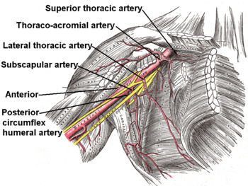
Axillary Artery Branches
The branches of the axillary artery include the following 6:
Axillary Artery Cannulation
Axillary artery cannulation is a possible way to establish cardiopulmonary bypass at the time of an ascending aorta and aortic arch surgery. A Dacron graft is used for the purpose, due to various potential benefits. During cannulation, axillary artery exposure is achieved through an incision of 6 to 10 cm atop the deltopectoral groove.
It is primarily indicated during operation including the aortic arch and the aortic root, once the pathology forbids a standard ascending aorta cannulation. Cannulation of the axillary artery can be used in:
When compared to the cannulation of a standard ascending aorta, that of Axillary artery has been found to reduce cerebral emboli. Axillary Arterial Line PlacementArterial line placement happens to be a common process in different critical care environments. Surgeons can place arterial lines in multiple arteries, which include the axillary, radial, dorsalis pedis, ulnar, femoral, posterior tibial and brachial arteries. Axillary arterial line placement is generally done by using anatomical landmarks or under ultrasound guidance.
Axillary Artery Injury and Problems
Axillary Artery Injury is slightly commoner than injuries involving the subclavian arteries, and account for around 5 – 10% of all arterial injury cases. It can be caused and associated to various issues, such as:
Patients may exhibit various symptoms, such as:
Commonly, associated trauma can include the axillary vein, which can then lead to injury to the branches or cords of the brachial plexus. Cases involving axillary artery and vein are common. Axillary Artery Aneurysms are harmful but rare lesions that put the upper extremities under risk with neurologic and vascular compromise. The majority of these lesions can be effectively treated with vascular grafting and surgical excision. Surgical cure is necessary for preventing the risks of ischemia and thromboembolism, which can consequently result in gangrene of the impacted extremity and its amputation. Naturally, operative treatment and management should not be delayed. Axillary Artery Pictures
Look at the following Axillary artery diagram and images to understand its anatomy, and the Axillary artery mnemonic.

|

|
|