|
|
Adductor Canal
Adductor Canal Definition
The adductor canal acts like a passage from the structures that move between the posterior leg and the anterior thigh. It is a tunnel that is conical and narrow in shape, and is situated in the thigh. This structure is 15 cm in length, and extends to the adductor hiatus located in the adductor magnus from the femoral triangle apex.
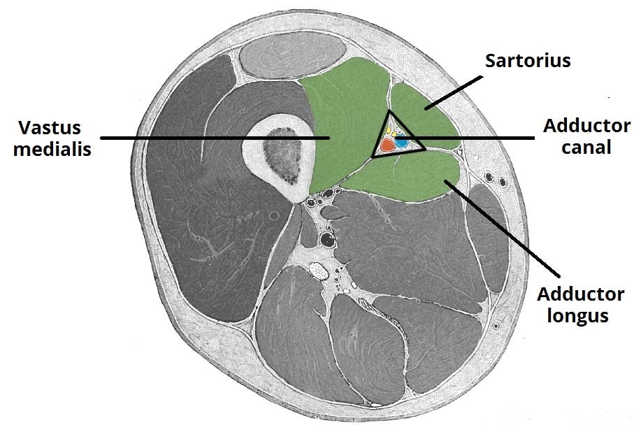
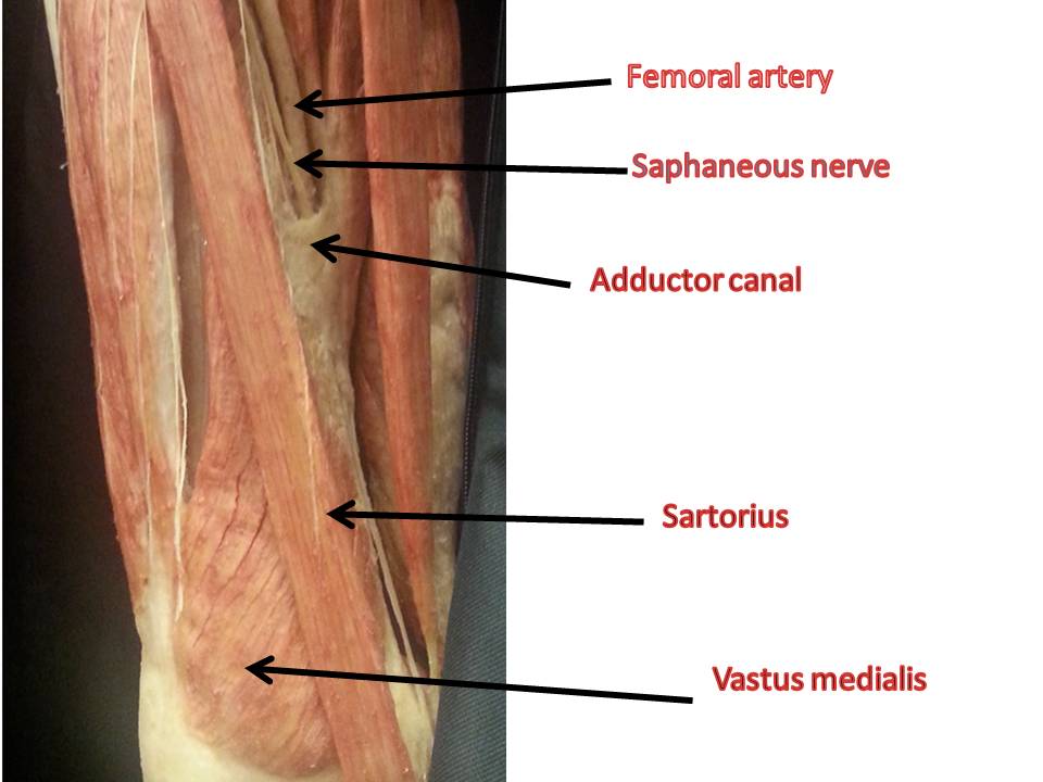
Muscular structures border the canal on various ends. There is Sartorius on the anterior end, the Vastus medialis on the lateral end and the Adductor magnus and Adductor longus on the posterior end. The adductor hiatus, a gap existing between the adductor magnus hamstring attachments and the adductor, marks the apex of the canal. This aponeurotic tunnel is also referred to as Subsartorial canal or Hunter’s canal. In Latin, it is known by the name ‘Canalis adductorius’. It is referred to as Canal des adducteurs in French. Adductor Canal Contents
The contents of Adductor Canal include:
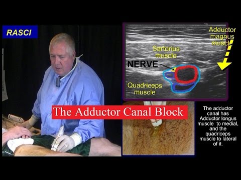
Adductor Canal Block
The adductor canal block is an innovative local anesthesia procedure for major knee surgery, which has come up in the last few years.
With this technique, there is fast analgesia onset that is similar to traditional femoral nerve blocks. However, the benefit with femoral blocks is the fact that the quadriceps muscles are spared. Naturally, no motor weakness arises in the leg as a consequence. With adductor canal blocks, there is superior pain management with better muscular strength. This can help in early ambulation after operation as well as rehabilitation. It can assist patients in satisfying discharge criteria much faster. The technique involves administering local anesthetic to the adductor canal for obstructing the saphenous nerve in isolation. The anesthetic may also be administered along with the nerve that moves to the vastus medialis. One can use the block to administer sensory anaesthesia for the processes that involve the lower leg, knee, femur and distal thigh on the medial side. The femoral artery and Sartorius are used as prominent marks to spot the saphenous nerve. Adductor Canal Compression Syndrome
Adductor Canal Compression Syndrome is a condition marked by neurovascular bundle entrapment in the adductor canal. This is a rare disorder that generally arises due to hypertrophy of vastus medialis and other muscles in the surrounding region. The condition is most common in young men, who can exhibit signs of claudication due to neurological symptoms arising due to saphenous nerve or more commonly, due to femoral artery occlusion.
Adductor Canal Pictures
The following Adductor Canal images and diagrams will help you to understand the location of Adductor Canal, as well as how AC Block is performed.
|

|
|